A Highly Relative Concept of Biological Normality

We all have that image in our heads—admittedly a bit simplistic—of the human skeleton we were shown in school. 206 bones, neatly arranged, a perfectly oiled mechanism that’s identical for everyone. It’s reassuring, isn’t it? Except that reality is much more… unpredictable. I often find myself thinking about just how much variation our bodies can tolerate—variations we can’t even imagine. In fact, some people retain bone structures that date all the way back to their embryonic development. They live with them, often without the slightest pain and without any visible external signs, sometimes well into old age.
This forces us to rethink our understanding of what constitutes biological “normality.” The number of bones, which seemed set in stone as an absolute truth, ultimately rests more on a statistical average than on a universal reality applicable to each of us. In some adults, small extra bones persist, unnoticed. These are direct remnants of our life in utero. These anatomical variations, long ignored or considered trivial, seriously challenge the idea of a body that is standardized like a machine rolling off an assembly line.
When Growth Leaves Its Mark: The Example of the Foot
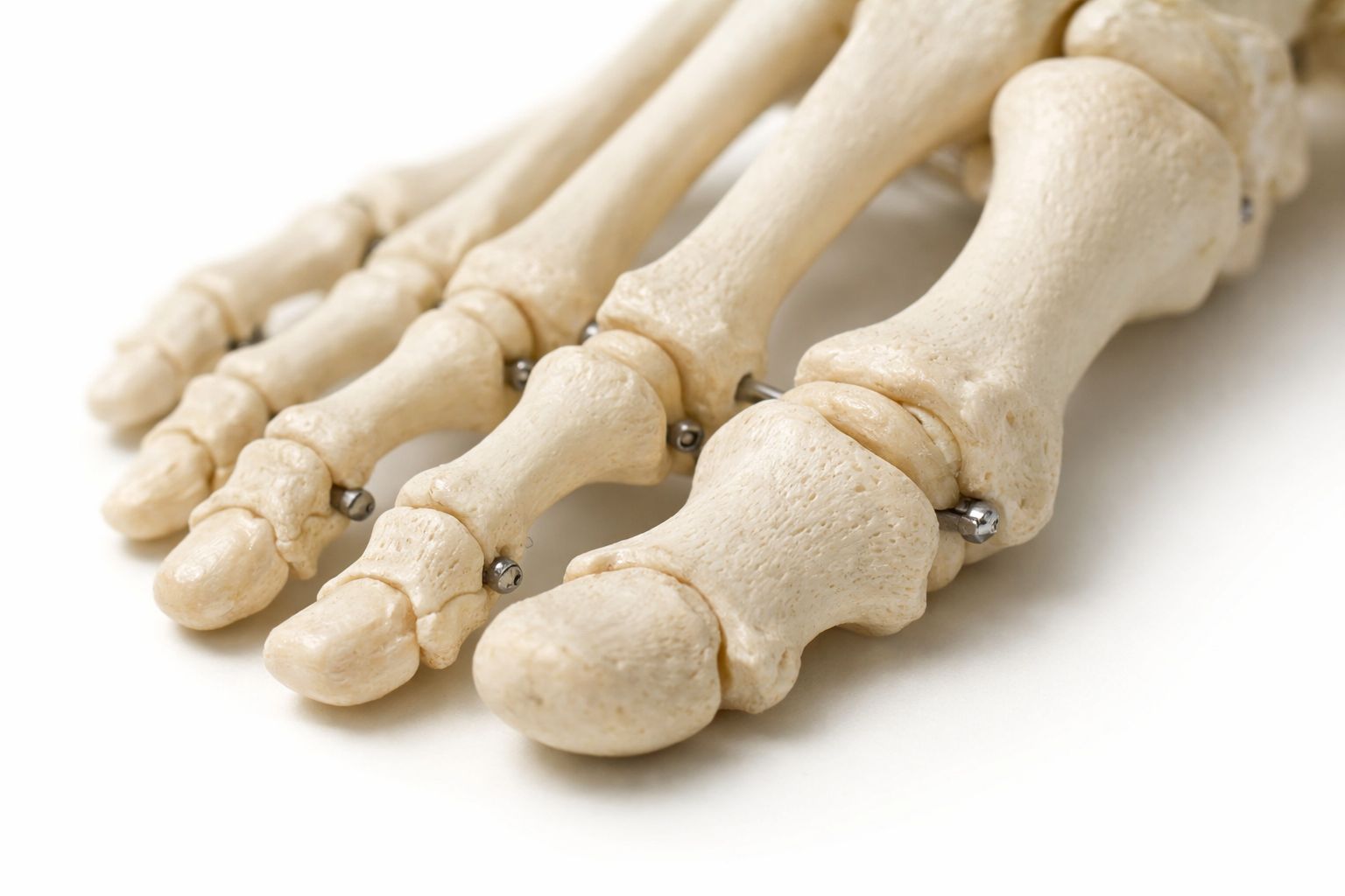
We must remember where we come from. At birth, a baby’s body is a sort of complex puzzle with far more bones than an adult has—sometimes nearly 300. That’s enormous, when you think about it. As we grow, nature generally streamlines the process: many ossification centers eventually fuse together gradually to form larger, stronger structures. But nature isn’t always as precise as a Swiss watch. In some people, this process remains partially incomplete. It leaves behind isolated bone fragments, like little reminders of the past.
I want to make it clear that these variations are neither a disease nor a rare, frightening anomaly. They simply reflect a different trajectory—a detour in bone development. Anatomists—those explorers of the human body—refer to these as secondary ossification centers, which, for one reason or another, never fused with the main bone. The phenomenon is actually quite common: it affects the feet just as much as the hips or even the cervical spine.
In fact, to give you an idea of the scale of the phenomenon, a very rigorous study published in the journal Scientific Reports in 2024 provided some statistics on the matter. Based on a meticulous analysis of Portuguese skeletal collections, the researchers showed that approximately 18% of individuals had at least one accessory bone in the foot. That’s nearly one in five people! This frequency is quite remarkable and reminds us that the human skeleton tolerates a great deal of variability without it necessarily affecting its day-to-day function.
Invisible “ghosts” often mistaken for fractures
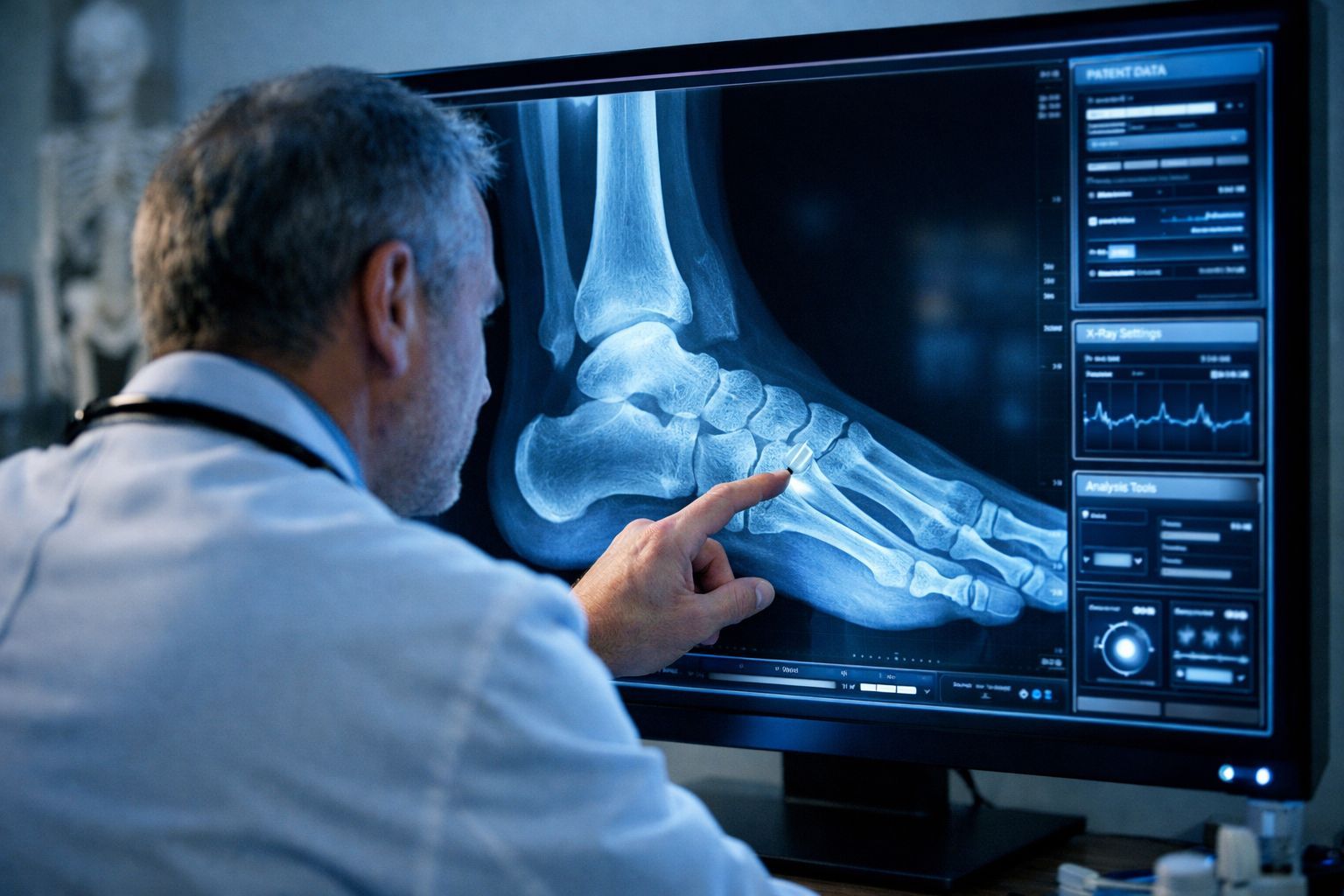
The most surprising thing about all this is the silence. In the vast majority of cases, these extra bones go completely unnoticed. They go about their business, so to speak, without causing any pain, functional discomfort, or even a visible change in morphology. You won’t spot a suspicious bump on your foot when you step out of the shower. They’re most often discovered purely by chance, during an X-ray or MRI performed for a completely unrelated reason. It’s often a complete surprise to the radiologist.
This inconspicuous nature explains why they have historically been underestimated. The catch is that even when they finally appear on a medical image, these accessory bones can cause confusion. They are sometimes mistaken—wrongly—for fragments of an old fracture that had been forgotten, or for age-related calcifications. According to observations by clinicians reported by LiveScience, they frequently go unreported or are misinterpreted, even by experienced professionals. To err is human, after all.
When Anatomy Speaks: Hip, Neck, and Unexplained Pain
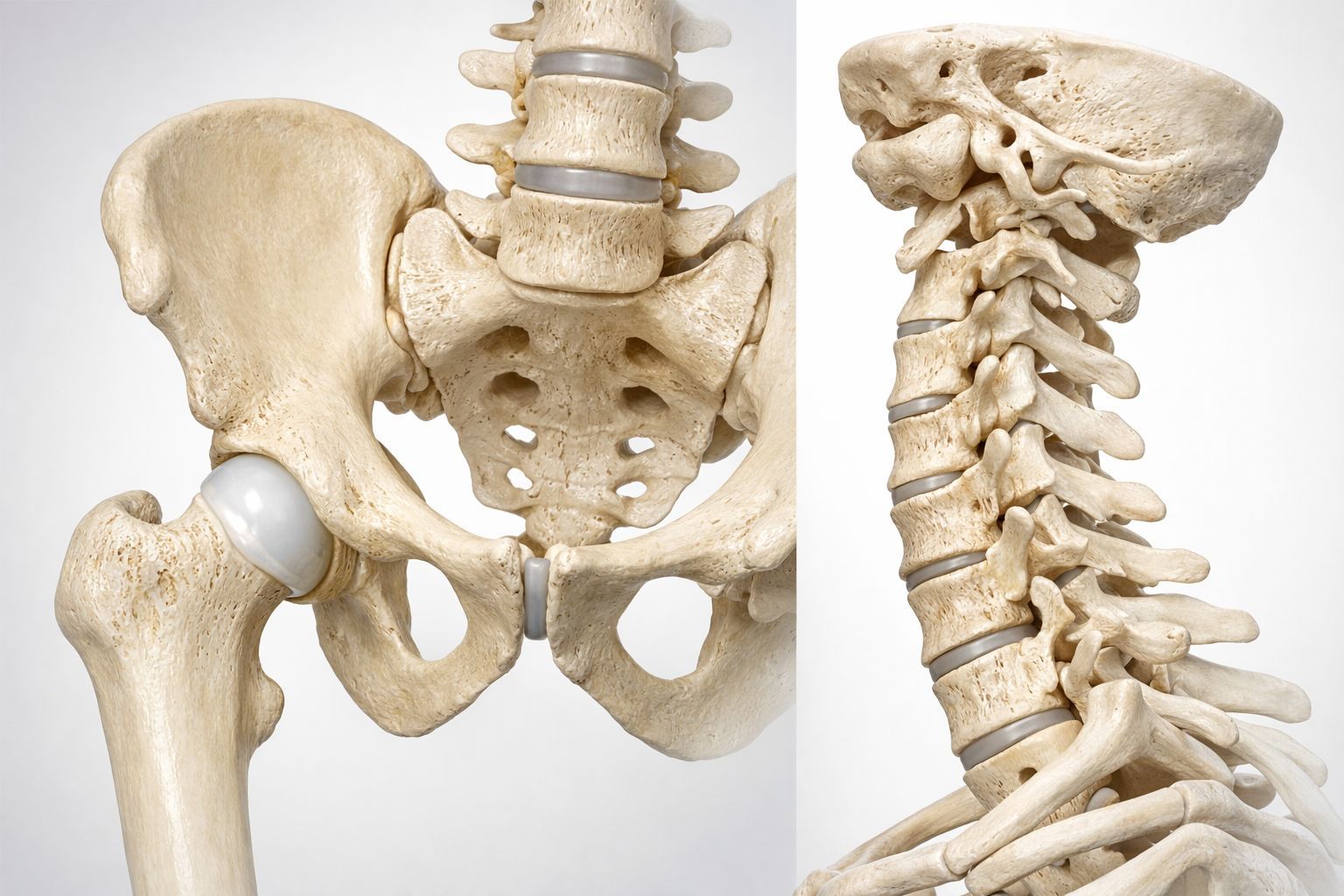
However, don’t assume this is always harmless. In certain specific contexts, these inconspicuous bones can become clinically significant—that is, they begin to cause problems. When they’re located in areas subject to high mechanical stress—think of the ankle or hip, which bear our weight—they can contribute to pain or limit joint range of motion. That’s when diagnosis becomes crucial.
A comprehensive review published very recently, in 2025, in the Bratislava Medical Journal examined this phenomenon, focusing on accessory bones of the hip, and in particular a certain acetabular bone. The authors show that this bone is present in less than 5% of the general population. That’s a small number, to be sure. But the figure rises among patients suffering from pain or joint conflicts. Its size and position directly influence symptoms and, in turn, the surgical decisions that will need to be made.
Another example—one that’s a bit unsettling but well-documented—involves the cervical ribs. These are supernumerary bones located at the base of the neck. According to the renowned Cleveland Clinic, up to 1% of people have one or two without even knowing it. The concern is that, in rare cases, they can compress nerves or blood vessels. The result? Pain and weakness in the arm. Correctly identifying them helps avoid inappropriate treatments and directs care toward the true cause of the problem. These situations remind us that these bones aren’t just museum curiosities; recognizing them plays a key role in understanding certain unexplained pains, particularly in athletes.
Conclusion

So, what does this teach us? That our bodies are a living archive of our own development. These small extra bones—these “intruders” that are mostly harmless—are proof that biology is not a rigid science.
The next time you have a minor, unexplained ache or get an X-ray, keep in mind that you might be part of that 18% or that tiny 1% who are out of the ordinary. It’s pretty fascinating to think that we never fully know ourselves, isn’t it?
You might have "extra" bones without even knowing it (and it’s fascinating)
This content was created with the help of AI.