When Technology Mimics the Breath of Life

We often get the impression that science has an answer for everything, don’t we? And yet, when it comes to understanding what’s really going on inside our lungs when a disease strikes, researchers are often… how should I put it… a bit in the dark. It’s frustrating. Until now, we’ve had to make do with animal models or cell cultures in static Petri dishes—which, let’s be honest, are a far cry from the complexity of a real human body that breathes, moves, and lives. But hold on to your hats, because a team may have found the key.
Imagine a lung… on a microchip. Yes, you read that right. A groundbreaking micro-respiratory device has just been developed. It allows us to monitor, almost in real time, the battle between our lung cells and infectious agents. This is a breakthrough that could completely revolutionize our fight against scourges like tuberculosis.
A technical feat: recreating life on a chip

So, how does this work? It’s not science fiction. Researchers at the renowned Francis Crick Institute, in collaboration with the biotech company AlveoliX, have developed this little gem, which they’ve named iLoC. It’s quite fascinating when you think about it: they’ve managed to recreate a living, functional, and immunocompetent alveolar environment. The secret? They use human stem cells harvested from a single donor. This is a crucial detail for genetic consistency.
Using a protocol with surgical precision, they’ve created the entire ecosystem needed: type I and II alveolar cells, endothelial cells, and even macrophages. All these cells share the same genetic makeup and are organized into a three-dimensional structure. It’s not just a random pile of cells; it forms an active respiratory barrier. And the craziest part is that it moves! The engineers designed microdevices that apply rhythmic stretches to the membrane, literally mimicking our breathing. Under the effect of this mechanical movement, the cells develop microvilli and increase their exchange surface area, exactly as in a real lung. According to the research, the system remains stable for two weeks, which gives us plenty of time to observe a lot of things.
What impresses me most is the level of detail. They’ve incorporated macrophages—those sentinels of our immune system—that patrol the barrier, secrete cytokines, and do their job of cleaning up. According to the work by Chak Hon Luk and his colleagues published in the journal Science Advances, this model even reveals an unexpected diversity of immune profiles, influenced by this mechanical stretching. In short, we’ve never achieved such realism in a simulation before.
A Deep Dive into the Heart of the Infection: Seeing the Invisible
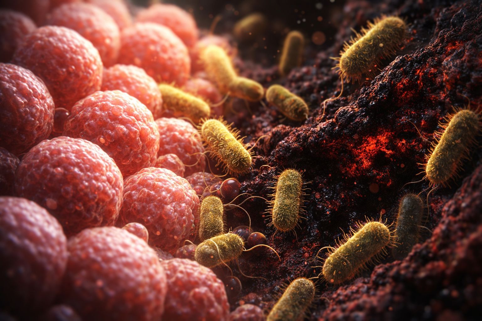
Well, having a miniature lung is all well and good, but is it actually useful? To find out, the researchers took a risky gamble: they exposed their device to Mycobacterium tuberculosis, the bacterium responsible for tuberculosis. They introduced it via the apical route to simulate an inhalation scenario, as if one were breathing in the bacteria on the street. And then… the show begins.
Within the first few hours—a stage that had never been observed before—the infection targets macrophages and alveolar cells. Endothelial cells, on the other hand, seem to be spared at first. It’s both fascinating and frightening. The bacterium infects the cells but doesn’t replicate right away. It bides its time. After a few days, necrotic foci begin to appear. Dead macrophages clump together, surrounded by cells that are still alive. On the fifth day, disaster strikes: the cellular barrier breaks down and alveolar function disappears completely. It’s brutal, but it’s exactly what we needed to understand.
The experiment didn’t stop there. They also tested a genetically modified version in which the macrophages were deprived of the ATG14 protein, which is involved in autophagy (cellular cleanup). The result? An increase in cell death, but strangely, the number of bacteria did not rise. Basically, without autophagy, our defenses collapse without the infection even needing to multiply further. This is a crucial finding.
Conclusion: Hope for the Future

Ultimately, what do we take away from all this? This iLoC model isn’t just a laboratory feat. It gives us access to the very earliest hours of a lung infection—a window of opportunity that had completely eluded us until now. It’s a bit like finally turning on the light in a dark room.
By adapting stem cells to specific genetic profiles, we can begin to envision truly personalized medicine. Imagine: we could predict a specific patient’s immune response or test the effectiveness of a targeted antibiotic before even administering it. This may be the beginning of the end for certain respiratory diseases that have been ruining our lives for far too long. Fingers crossed, right?
This little piece of breathable plastic could very well change the history of tuberculosis
This content was created with the help of AI.